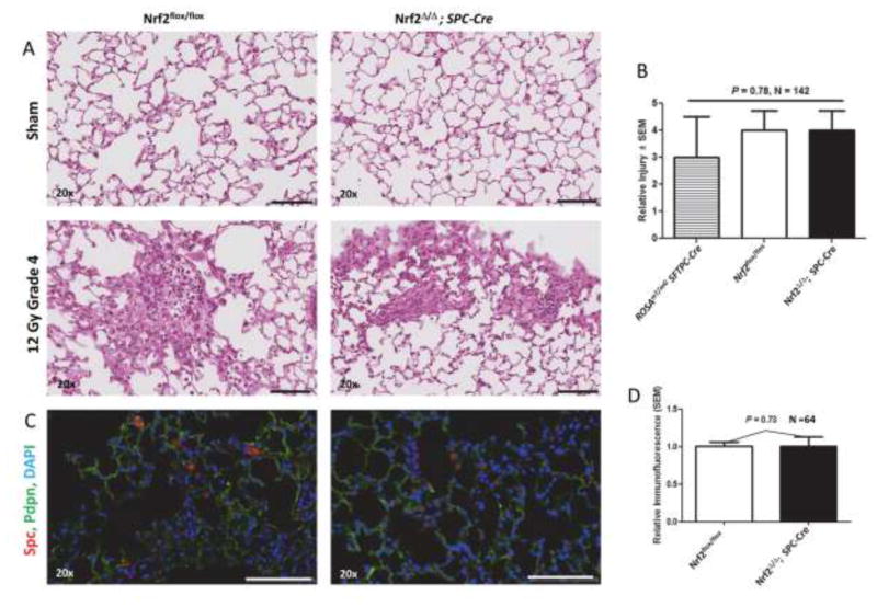
Figure 3.
Tissue remodeling and Spc immunofluorescence in sham (0 Gy) and irradiated (12 Gy) lung of Nrf2flox/flox and Nrf2Δ/Δ; SPC-Cre mice 250 days after irradiation. A) Representative H&E stained sections. B) Quantification of tissue remodeling in irradiated ROSAmT/mG SFTPC-Cre, Nrf2flox/flox and Nrf2Δ/Δ; SPC-Cre mice. C) Representative sections immunostained for Spc (red) and Pdpn (green), and counter stained with DAPI, imaged using wide field whole slide scanner. microscopy. D) Quantification of relative Spc immunofluorescent cells per field corrected for DAPI staining. Black and white bars = 100 μm.

