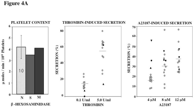
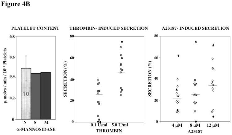
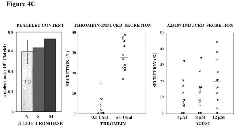
Figure 4. Platelet Content and Secretion of β-Hexosaminidase (A), α-Mannosidase (B), and β-Glucuronidase (C) in the propositus and his mother.
In each panel the bar diagram shows the platelet content of the acid hydrolase in the two patients and 10 normal subjects (N, mean ± SE). Platelet secretion is shown for each acid hydrolase in the propositus (filled circles), his mother (filled triangles, studied twice) and 10 healthy controls upon activation with thrombin and A23187.


