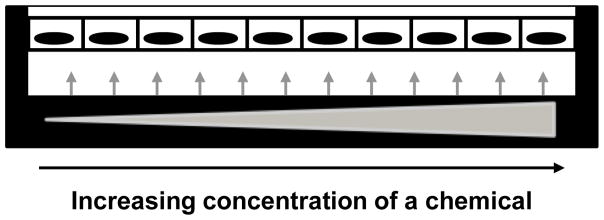
Figure 2. Applying a chemical gradient in cell culture.
A cell culture specimen receives a chemical at increasing concentrations (as displayed by the widening triangle and the arrows) depending on the location in the culture area. Here cells of an epithelium are represented by rectangles with nuclei drawn in black, in the culture platform. With such system, it is possible to create controlled heterogeneity of the presence of the chemical throughout the culture surface. It is also possible to identify concentration thresholds for the impact of the chemical on cells depending on a given variation in cell culture parameters.

