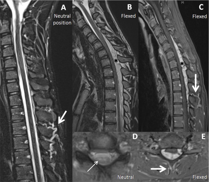
Figure 1.

Venous dilation on cervical MRI of a patient with Hirayama disease. Marked dilation of posterior soft tissue veins (thick arrows) detectable in neutral (A) and flexion position (C, E); the crescent-shaped dilation of the epidural venous plexus (asterisks) was detectable only in flexion position (B, C); snake-eyes sign (thin arrow) (D) (A, B and D: T2 sequences; C and E: contrast-enhanced T1 sequences).
