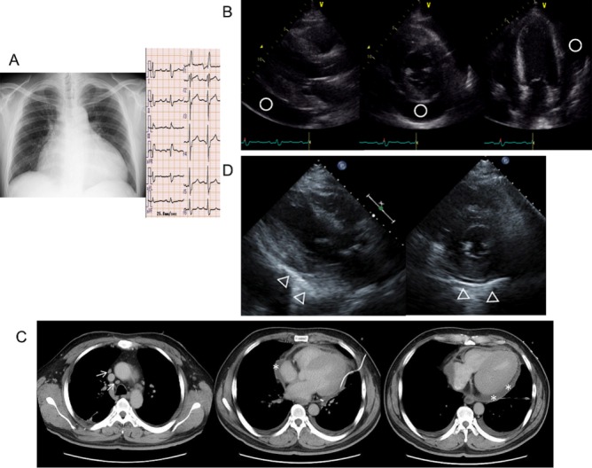
Figure 1.

(A) Chest radiography and ECG at admission chest radiography shows cardiomegaly (cardiothoracic ratio, 69.6), and ECG shows sinus tachycardia and complete right bundle branch block. (B) Transthoracic echocardiography at admission. Left ventricular ejection fraction is preserved (53.4%), and there is a large amount of pericardial effusion (○). (C) Chest CT scan after pericardial effusion drainage. There is swelling of the mediastinal lymph nodes (↑) and residual pericardial effusion (*). (D) Transthoracic echocardiography performed 35 days after pericardial effusion drainage. Pericardial effusion is not noted; however, the pericardium shows partial calcification (Δ), especially at the posterior and inferior sides of the left ventricle. Additionally, the surface of the heart has a rough appearance with diminished movements.
