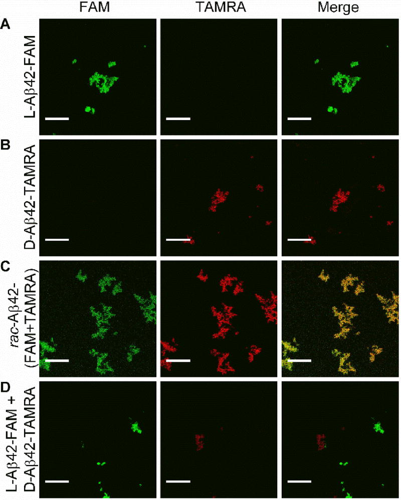
Figure 2.
Two-channel confocal microscopy imaging (Channel 1, left panel, monitors FAM: excitation at 476 nm, emission over 484–514 nm; Channel 2, middle panel, monitors TAMRA: excitation at 543 nm; emission over 630–690 nm; right panel: merging of Channels 1 and 2 allows to probe for co-localization of the fluorescent labels). L-Aβ42-FAM fibrils were fluorescentl in Channel 1, but not Channel 2 (A), whereas D-Aβ42-TAMRA fibrils were active via Channel 2 only (B). Fibrils grown from an equimolar mixture of L-Aβ42-FAM and D-Aβ42-TAMRA were fluorescently active in both Channel 1 and 2, with robust signal co-localization (C). In a control experiment (D), fibrils grown from either L-Aβ42-FAM or D-Aβ42-TAMRA were subsequently mixed and were fluorescently active either in Channel 1 or Channel 2, with very low co-localization. Scalebar: 20 µm.

