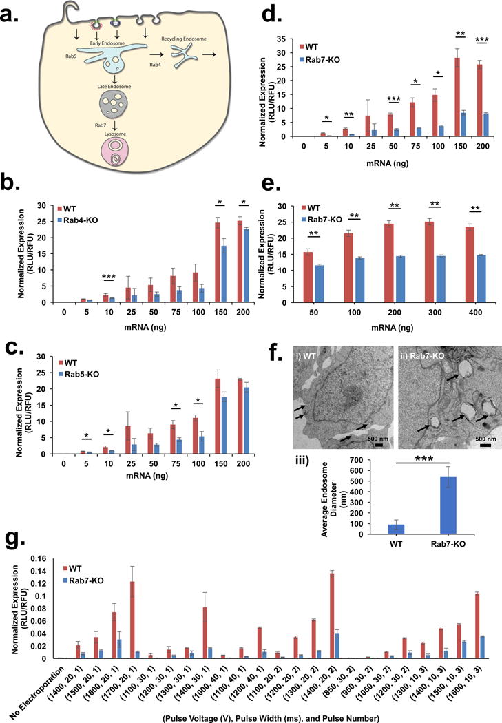
Figure 1. Late endosomes are essential for mRNA transfection.

(a) Schematic representation of Rab proteins localizing to various stages of endocytic process. (b–d) HAP1 cells with deletions of (b) Rab4A, (c) Rab5A, and (d) Rab7A were transfected with mRNA packaged inside lipoplexes and luciferase expression was compared to wild-type. (e) HAP1-Rab7-KO cells were transfected with LNPs at a range of mRNA doses and normalized luciferase expression was compared to wild-type (f) Electron micrographs of (i) HAP1-WT (scale bar = 500 nm) and (ii) HAP1-Rab7-KO cells (scale bar = 500 nm) showing enlarged endosomes in Rab7-KO cells indicated by arrows. (iii) Average size of endosomes (indicated by inset arrow) in HAP1-WT and HAP1-Rab7-KO, n = 4, mean ± SD. (g) Luciferase expression in HAP1-WT and HAP1-Rab7-KO cells transfected with free mRNA using electroporation at various pulse voltages, pulse widths and pulse numbers. All experiments were conducted with n = 3; mean ± SD, unless indicated otherwise. Statistical analysis of the data was assessed by Student’s t-test (0.05 ≥ *p > 0.01, 0.01 ≥ **p > 0.005, ***p ≤ 0.005).
