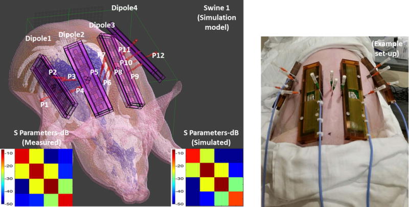
Figure 2.

Anatomical swine models are generated from CT images. Anatomy is segmented to fat, muscle, bone, skin and inner air. Thermal probes and dipole blocks were also segmented from the same images. S parameter matrices obtained from experiments and simulations are also shown.
