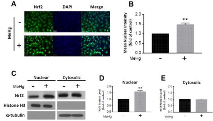
Figure 2.

Nrf2 translocates to the nucleus in response to MeHg exposure. Astrocytes were treated with 5 μM MeHg for 6 hours, and immunostained for Nrf2 (A). Nrf2 mean intensity in cell nuclei was quantified from four distinct images (B; n = 4). Further, Nrf2 protein level was measured in nuclear (C,D; n = 3) and cytosolic fractions (C,E; n = 3) separated by subcellular fractionation. Representative blots for nuclear and cytosolic Nrf2, histone H3, and α-tubulin are displayed (C). Nuclear and cytosolic fractions were normalized to histone H3 and α-tubulin, respectively. All data were normalized to untreated controls, and represent the mean ± SEM. **p < 0.01, scale bar = 60 μm
