
Fig. 4. Stg expression disrupts but does not block bristle development.
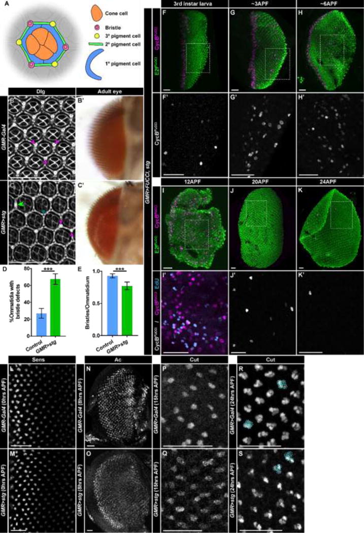
(A) Diagram of cell types in an ommatidium visible in apical sections of the retina at 48hrs APF. The morphological organization of the ommatidium allows identification of each cell type using membrane staining. (B,B′) Retinas from control (GMR-Gal4) flies. (B) Retina at 48hrs APF stained with antibodies recognizing Dlg, which marks cells membranes. Bristles (purple arrows) are normally located at alternating vertices of the hexagonal ommatidia, generating a regular pattern in the adult eye (B′). (C,C′) Retinas from GMR>stg flies. (C) In a retina stained with anti-Dlg at 48hrs APF, bristles are misplaced. Examples include bristles located at neighboring vertices (purple arrows), absent bristles (blue asterisk), and bristles located in between two vertices (green double arrow). Bristles are visible in adult eyes (C′). (D) Quantification of ommatidia per retina with bristle defects (less than or more than three bristles; bristles at neighboring vertices; bristles in between two vertices). Control is GMR-Gal4/CyO. p<0.0001; n>5 retinas, with >180 ommatidia counted/retina. (E) Quantification of average number of bristles per ommatidia for control (GMR-Gal4/CyO) and GMR>stg. p<0.001 n>5 retinas, with >180 ommatidia counted/retina. (F–K′) GMR>FUCCI, stg retinas expressing E2fFUCCI (green) and CycBFUCCI (F–K,I′, magenta; F′–G′,J′,K′ grey) analyzed at the indicated times after pupal formation (APF). (I′) Retina at 12APF stained with EdU (blue). Dotted boxes indicate areas of magnification in E′–J′. (L,M) Retinas at 0hrs APF stained with anti-Sens antibodies in (L) control and (M) GMR>stg. (N,O) Retinas at 8hrs APF stained with anti-Achaete/Ac antibodies in (N) control and (O) GMR>stg. (P,Q) Retinas at 15hrs APF stained with anti-Cut antibodies in (P) control and (Q) GMR>stg. (R,S) Retinas at 24hrs APF stained with anti-Sens antibodies in (R) control and (S) GMR>stg. Four cell Cut-expressing groups (circled) are present in both genotypes; groups that appear to have less than four cells have additional cells outside the plane. Scale bars = 20μM.