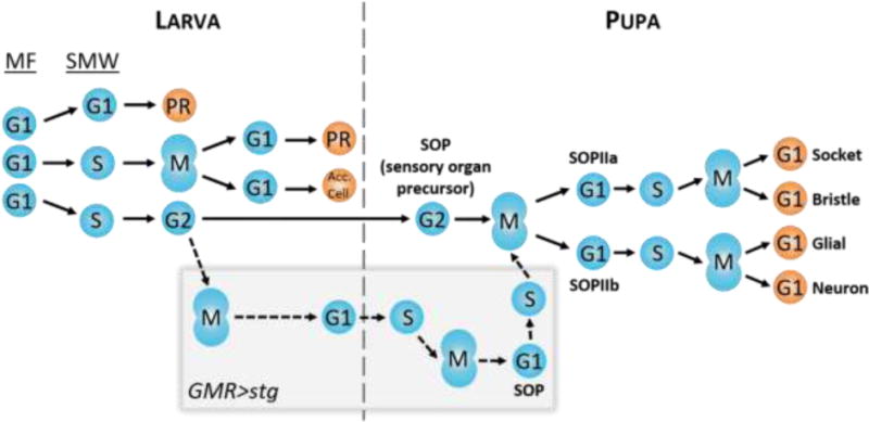
Fig. 5. Model of interommatidial development.

As the larva develops, the MF initiates cell cycle synchronization by promoting G1 arrest in all cells. These cells will subsequently take one of three paths: 1) arrest permanently in G1 and undergo differentiation into photoreceptors R2/3/4/5/8 (PR); 2) undergo S phase of the SMW, followed by mitosis, permanent arrest in G1, and differentiation into photoreceptors R1/7/6 and accessory cells (Acc. Cell; i.e. cone cells and pigment cells); 3) undergo S phase of the SMW and arrest in G2 for the remainder of larval development, followed by selection as an SOP during pupal development and two divisions to make up the four G1-arrested cells of the bristle. Prematurely driving cells through mitosis, such as with GMR>stg (gray box), does not prevent SOP selection and bristle differentiation (dotted lines). Differential timing of SOP divisions between wild type and GMR>stg (not shown in this model) may contribute to bristle positioning defects observed in GMR>stg adult eyes.
