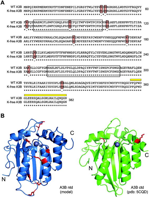
Fig 1. Lysine residues in human APOBEC3B.

(A) Amino acid alignment of wild-type and K-free A3B. Lysine to arginine substitutions are highlighted with red boxes, N-terminal and C-terminal zinc-coordinating domains by open boxes, and 5210-87-13 mAb epitope by a thick yellow line.
(B) Ribbon structures of A3Bntd (model) and A3Bctd (pdb: 5CQD) with lysines colored red.
