
Fig. 5. Nuclear localization of APOBEC3B does not require lysines.
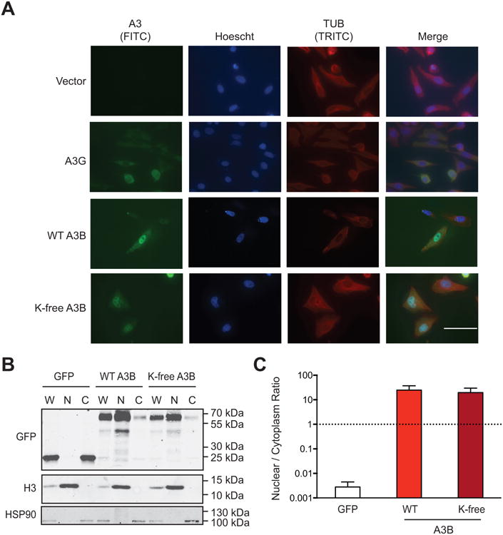
(A) Representative images of HeLa cells expressing an empty vector, A3G (untagged), wild-type A3B (untagged), or K-free A3B (untagged). The rabbit mAb 5210-87-13 was used to detect each A3 protein through a nearly identical C-terminal epitope (anti-rabbit IgG-FITC as secondary). 100 μM scale applies to all images.
(B) Immunoblot of 293T whole cell (W), nuclear (N), and cytoplasmic (C) extracts expressing eGFP, wild-type A3B-eGFP, or K-free A3B-eGFP. Fractionation controls are anti-Histone H3 for nuclear components, and anti-HSP90 for cytoplasmic components.
(C) Quantification of the fractionation data from panel B and 2 biologically independent experiments (not shown). Each histogram bar reports the mean +/- SEM nuclear to cytoplasmic ratio for the indicated constructs.