
FIGURE 2.
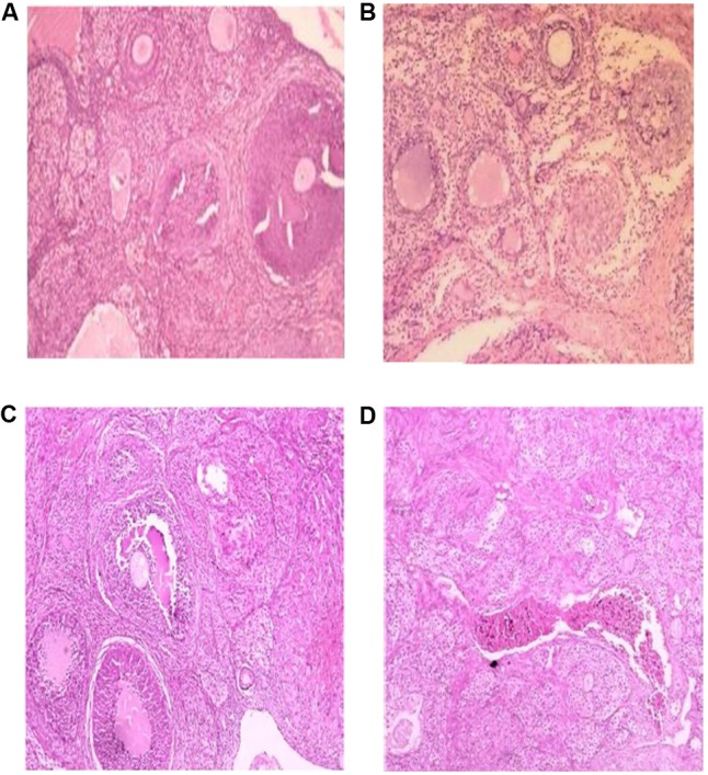
Effect of AEGD on the ovary histological analysis of the treated rats (hematoxylin–eosin, 100×). (A) Section of (vehicle control, distilled water) rat ovary showing normal stroma with primary and secondary developing follicles and matured graafian follicle. (B) Section of (chronic restraint stress) rat ovary showing hyperchromatic nucleus, multiple follicular cysts and atretic follicles and corpus fibrosum. (C) Section of (chronic restraint + G. dalenii 7.5 mg/kg) rat ovary showing only few developing follicles. (D) Section of (chronic restraint stress + G. dalenii 15 mg/kg) rat ovary showing most follicles with normal follicular development and intact corpus luteum.