
Figure 1.
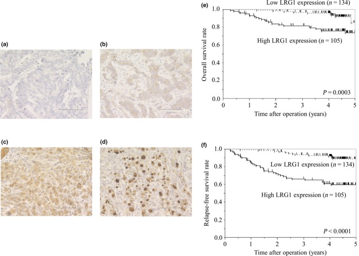
(a–d) Leucine‐rich α2‐glycoprotein‐1 (LRG1) immunohistochemical staining in gastric cancer specimens at ×200 magnification. Staining results are shown at: score 0, no stained cancer cells in the section (a); score 1, ≤10% of cells immunoreactive to LRG1 (b); score 2, >10% of cells immunoreactive to LRG1 (c); and score 3, >10% of cells immunoreactive to LRG1 and also found both in cytoplasm and nuclei (d). Bar = 100 μm. (e,f) Kaplan–Meier analyses of overall survival (e) and relapse‐free survival (f), based on LRG1 expression.