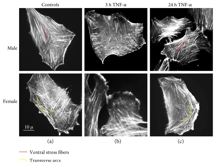
Figure 2.
Effect of TNF-α on actin filament organization. Static cytometry analysis of actin cytoskeleton in cells stained with fluorescein—phalloidin. Numerous ventral stress fibers in control cells from males and transverse arcs in control cells from females (a) are detectable; stress fiber loss and intense lamellipodia formation are visible 3 hours after TNF-α treatment (b) and reappearance of stress fibers after 24 hours of TNF-α treatment (c).

