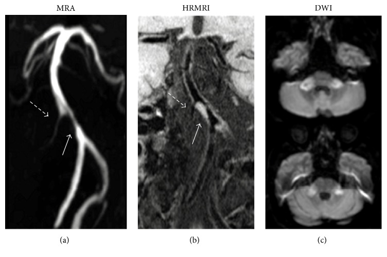
Figure 3.
Images from a 65-year-old man with subacute bilateral brachium pontis infarction. (a) MRA shows stenosis of BA (solid arrow) and right anterior inferior cerebellar artery (dashed arrow). (b) T1WI-SPACE images (coronal) show plaque on BA (solid arrow), right anterior inferior cerebellar artery (dashed arrow), and anatomical relationship between orifices of right anterior inferior cerebellar artery and plaque.

