Figure 8.
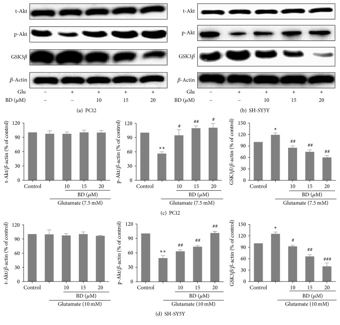
Effects of biatractylolide on Akt and Gsk3β expressions of PC12 and SH-SY5Y cells induced by glutamate. (a)-(b) represent protein characterization of Akt and Gsk3β after PC12 and SH-SY5Y cells were treated with various concentrations of biatractylolide for 30 min before glutamate treatment for 24 hours. Blots were also probed for β-actin as loading controls. (c)-(d) showed the ratio of different proteins to β-actin was calculated by the band density of each cell line using Image J software. ∗p < 0.05 and ∗∗p < 0.01 versus control group; #p < 0.05, ##p < 0.01, and ###p < 0.005 versus model group.

