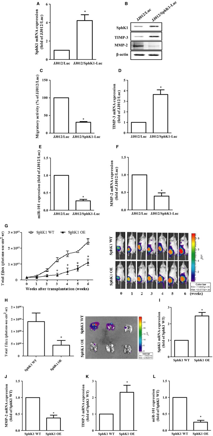
Figure 5.

Overexpression of SphK1 decreases cell migration and chondrosarcoma metastasis in vitro and in vivo. The constitutively expressed pLenti CMV V5‐Luc JJ012 cells were transfected with pCMV6 plasmid alone (JJ012/Luc) or harboring with human SphK1 ORF cDNA (JJ012/Sphk1‐Luc), followed by the determination of the protein (B) and mRNA expressions of SphkK1 (A), TIMP‐3 (D), MMP‐2 (F), as well as miR‐101 expression (E) and the migratory activity (C) by immunoblotting, real‐time PCR, and the transwell analyses, respectively. (G) JJ012/Luc or JJ012/SphK1‐Luc cells representing the wild‐type or overexpression of SphK1 were injected into the lateral tail vein of severe combined immunodeficient mice, and the development of lung metastasis was monitored by bioluminescence imaging at the time intervals. These images were then quantified (photons/s of lung region). After six weeks, these mice were humanely sacrificed, and the lung tissues were excised, photographed, and quantified (H). The mRNA expressions of SphK1 (I), MMP‐2 (J), and TIMP‐3 (K), as well as miR‐101 (L) on these tumors, were then assessed by real‐time PCR analysis. Cells without overexpression of SphK1 were used as the control (set to 1 or 100), and data were shown as multiples of that. The results are expressed as mean ± SEM. *P < 0.05 compared with control (n ≧ 3). SphK1, sphingosine kinase 1; ORF, open reading frame; WT, wild‐type; OE, overexpression.
