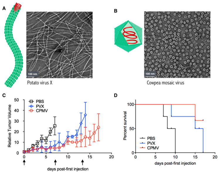
Figure 1.

(A,B) Schematic and transmission electron micrographs of (A) PVX and (B) CPMV. (C,D) Tumor treatment study. Tumors were induced with an intradermal injection of 125 000 cells/mouse. Mice (n = 3) were treated with 100 μg of PVX or CPMV (or PBS control) once weekly, starting ∼8 days post induction when tumors measured <100 mm3. Arrows indicate injection days; mice were sacrificed when tumor volumes reached 1000 mm3. (C) Tumor growth curves shown as relative tumor volume. (D) Survival rates of treated mice.
