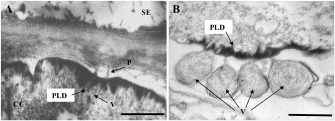
FIGURE 2.

Transmission electron micrographs showing LIYV-induced conical plasmalemma deposits (PLDs). (A) Shows PLDs located at the internal side in a companion cell (CC) of a LIYV-infected Nicotiana benthamiana leaf, associated with LIYV virions (V) and plasmodesmata (P) [image is modified from Medina et al. (2003) with permission of John Wiley and Sons]. (B) Shows PLDs in a LIYV-infected N. benthamiana protoplast, sacks of LIYV virions (V) are external to the plasmalemma directly adjacent to abundant PLDs [image is modified from Kiss et al. (2013) under the CC BY License]. Labeling is CC, companion cell; P, plasmodesmata; PLD, plasmalemma deposit; SE, sieve element; V, LIYV virions.
