Figure 2.

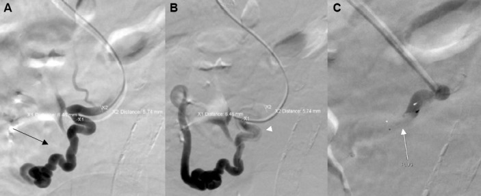
(A) Portogram showing single dominant duodenal varix (black arrow). (B) Premicrovascular plug deployment selective venogram (white arrowhead). (C) Post plug deployment over the wire venogram demonstrating instant occlusion (white arrow).

(A) Portogram showing single dominant duodenal varix (black arrow). (B) Premicrovascular plug deployment selective venogram (white arrowhead). (C) Post plug deployment over the wire venogram demonstrating instant occlusion (white arrow).