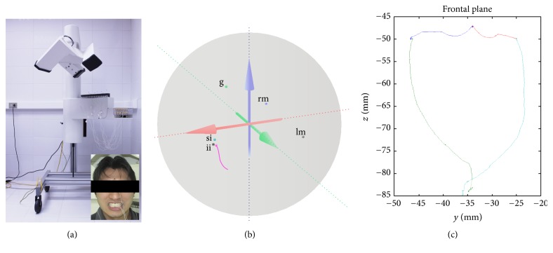
Figure 1.
(a) AG501 3D Electromagnetic Articulograph (EMA) and properly positioned sensors: 2 mastoids (reference), 1 glabellar (reference), 1 upper incisor (reference), and 1 lower incisor (movement); (b) spherical volume of analysis (30 cm of diameter) showing the sensor positioned in volunteers (g: glabellar; rm: right mastoid; lm: left mastoid; si: superior incisor; ii∗: inferior incisor/movement) and an opening trajectory (pink line); (c) trajectory obtained from border mandibular movements in the frontal plane.

