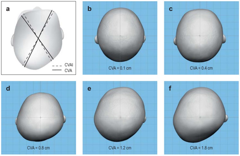
Figure 2.
a) Schematic depiction of cephalometric measurements (see also eFigure 1). The solid line shows the measurement of the cranial vault asymmetry (CVA) according to Moss and Mortenson et al. (e5, e6), based on the difference between the largest and smallest diagonal diameter. The dotted line shows the measurement of the cranial vault asymmetry index (CVAI) according to Loveday et al. (e7), based on two diagonals that are both angled at 30° to the mid-sagittal plane. b–f) Stereophotogrammetric images (top view) with differing cranial vault asymmetry (CVA). Even though the image cannot visualize all clinical signs, compensatory prominence of the forehead and compensatory widening of the skull with increasing degrees of severity are clearly recognizable.

