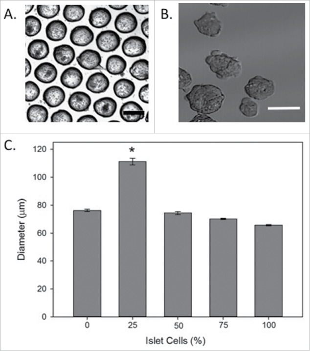
Figure 1.

Formation of Spheroids. (A) Cells were loaded into micromold plates and began to cluster into spheroids within 24 hours. (B) By 3–5 days, mature spheroids were removed from the micromolds and used for testing. The image shows examples of pure MSC spheroids, termed 0% spheroids. Scale bar = 100μm for both images. (C) The average diameter of spheroids in each group at day 1 was below 100µm with the exception of the 25% islet group. For diameter measurements, spheroids from 4 independent trials were measured with a total of 135–285 spheroids/group. * indicates a significant increase in spheroid diameter, p < 0.05.
