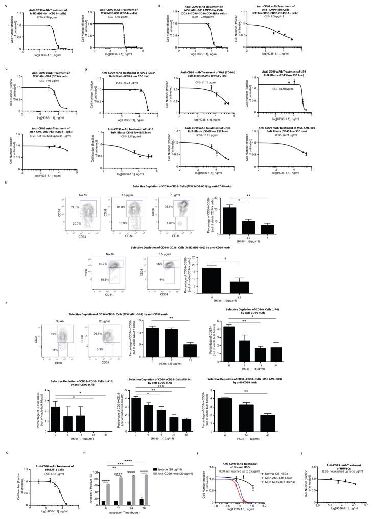
Fig. 3. An anti-CD99 monoclonal antibody (mAb) is cytotoxic to MDS and AML cells.
(A) Incubation of purified CD34+ cells from MSK MDS-001 and MDS-002 with anti-CD99 mAb (clone H036-1.1) for 48 hours led to a significant decrease in cell number. Similar results were obtained with (B) CD34+CD38−CD90−CD45RA+ LMPP-like cells from AML specimens MSK AML-001 and UP31, as well as (C) CD34+ cells from MSK AML-004. CD34+ cells from the BCR-ABL positive AML MSK AML-005 were incubated with anti-CD99 mAb (H036-1.1) for 48 hours. The IC50 was not reached using mAb concentrations up to 35 μg/ml. (D) Incubation of bulk blasts (CD45[low]SSC[low]) purified from UP32, UA8, UP4, UA16, UP34, and MSK AML-003 with anti-CD99 mAb (clone H036-1.1) for 48 hours led to a significant decrease in cell number. (E) At the end of the incubation of MSK MDS-001 and MSK MDS-002 with anti-CD99 mAb, CD34 and CD38 expression was measured on remaining viable cells, demonstrating selective depletion of CD34+CD38− cells. Representative FACS-plots are shown. *P<0.05, **P<0.01 (unpaired t-test). (F) At the end of incubation of MSK AML-004, UP4, UA16, UP34, and MSK AML-003 with anti-CD99 mAb, CD34 and CD38 expression was measured on remaining viable cells, demonstrating selective depletion of CD34+CD38− cells (or CD34+ cells in UP4). Representative FACS-plots are shown here and in Supp. Fig. 12. *P<0.05, **P<0.01 (unpaired t-test). (G) MOLM13 cells were incubated with anti-CD99 mAb (H036-1.1) for 48 hours, leading to a marked decrease in cell number. (H) Incubation of MOLM13 cells with anti-CD99 mAb (H036-1.1, 20 μg/ml) led to induction of apoptosis over the course of 36 hours. **P<0.01, ***P<0.001, ****P<0.0001 (unpaired t-test). (I) 700 HSCs (LN CD34+CD38−CD90+CD45RA−) purified from CB were incubated with anti-CD99 mAb (H036-1.1) for 48 hours. The IC50 was not reached using mAb concentrations up to 35 μg/ml. Juxtaposed for comparison is the sensitivity of MSK MDS-001 and MSK AML-001 to anti-CD99 mAb as shown in panels A and B. (J) Similar results were obtained when human umbilical vein endothelial cells (HUVECs) were incubated with anti-CD99 mAb (H036-1.1) for 48 hours. For panels A–J, error bars represent ±SEM of biological triplicates.

