
Figure 1.
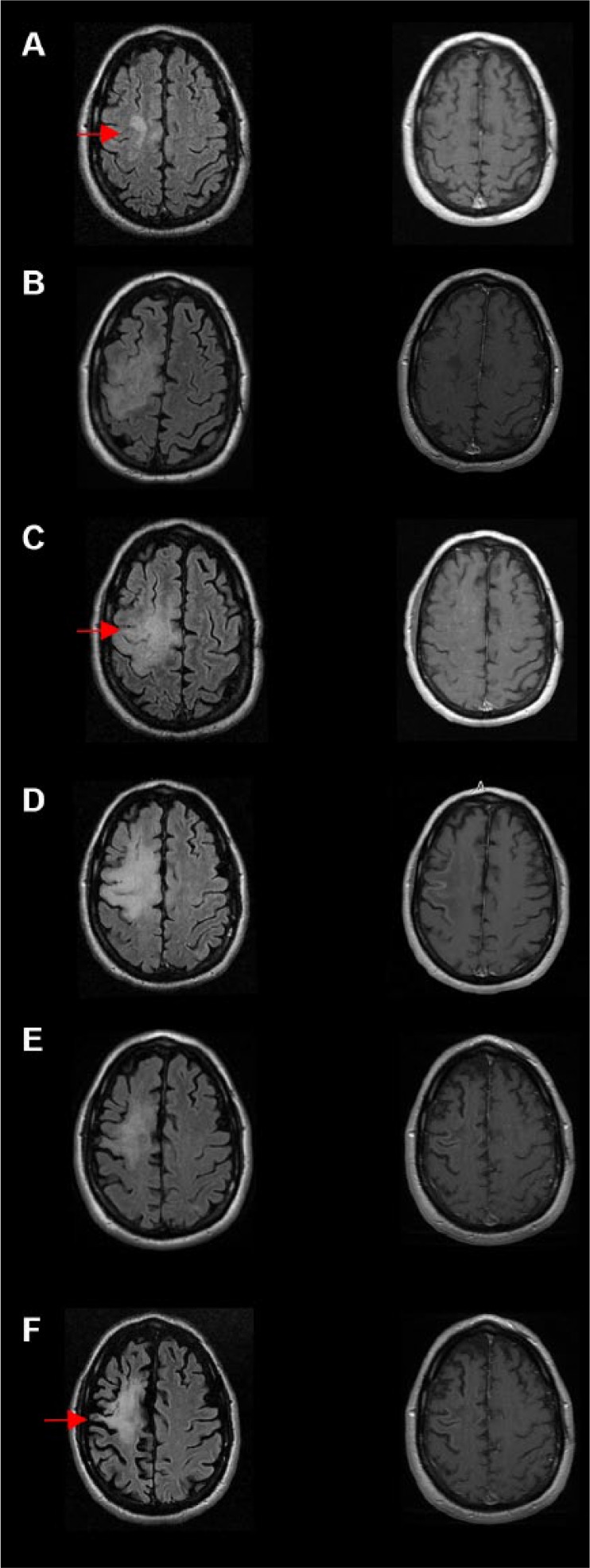
MRI progression of progressive multifocal leukoencephalopathy (PML) and immune reconstitution inflammatory syndrome (IRIS). (A) Initial imaging suspicious for PML. A new confluent hyperintensity was shown in the right superior frontal cortical and subcortical white matter region (arrow). This was confirmed to be PML with CSF JCV studies. (B) The patient presented 2 months later with worsened symptoms, and MRI showed worsened and enlargement of the lesion on T2 FLAIR (arrow; left column B, C, D), with patchy enhancement on T1 post contrast (right column B, C, D). The worsening clinical picture represents acute IRIS. Images C, D, and E show the progression of acute PML-IRIS lesions 2.5 months, 4 months, and 7 months from the diagnosis. (F) Decreased hyperintensity with increasingly apparent brain atrophy (arrow) was seen 14 months after the diagnosis. Left-side column: T2 FLAIR sequence; Right-side column: T1-post contrast.