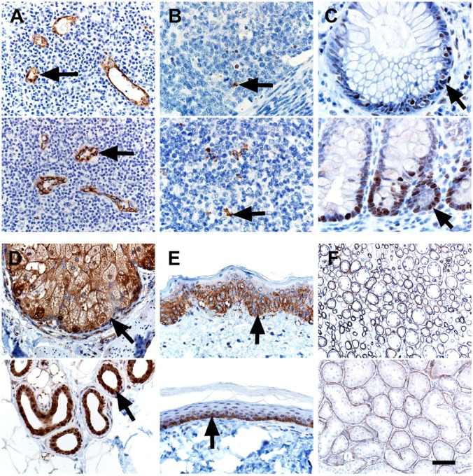
Figure 2.
IHC of human (top panel) and pig (bottom panel) tissues. (A) vWF immunostaining was localized to endothelial cells (arrows) of vessels. (B) Cleaved Caspase-3 immunostaining was localized to apoptotic cells (arrows) and debris in lymphoid tissues. (C) PCNA immunostaining was localized to nuclei of proliferating epithelial cells (arrows) in the crypts of the colon. (D) EMA10 immunostaining was localized to adnexal glands (arrows) of the skin. (E) Glut1 immunostaining was localized to the lower, basal-oriented cells (arrows) of the epidermis. (F) Laminin immunostaining was localized to the tubular basement membranes in the kidney. Scale bar = 26 (B, C), 40 (A, D, E), and 80 (F) µm. Abbreviations: vWF, von Willebrand Factor; PCNA, proliferating cell nuclear antigen; EMA, epithelial membrane antigen; Glut1, glucose transporter 1.

