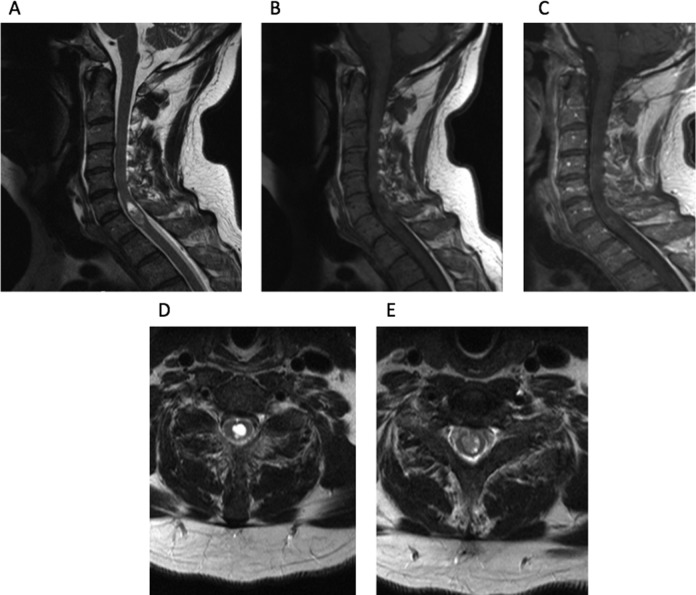
Figure 1.
(A) Sagittal T2-weighted MRI reveals lesion centered at C5-7, with an associated fluid collection at the most rostral part of the lesion. (B) Sagittal T1-weighted MRI of lesion. (C) Sagittal contrast-enhanced MRI reveals scant rim enhancement. (D) Axial T2-weighted MRI centered on the fluid collection. (E) Axial T2-weighted MRI at more caudal part of the lesion.

