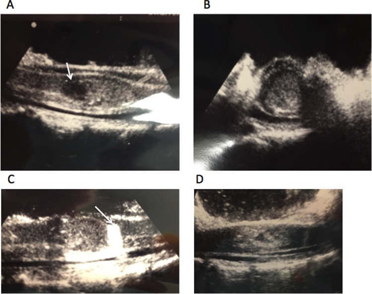
Figure 2.
Intraoperative ultrasound of spinal cord after laminectomy reveals lesion. (A) Fluid collection can be seen at the left (white arrow). (B) Looking at axial perspective, one can see lesion encompassing most of the spinal cord. (C) Using a piece of Gelfoam (white arrow), one is able to make sure that most caudal aspect of lesion is exposed during intramedullary dissection. (D) Ultrasound after resection of lesion reveals resolution of mass effect.

