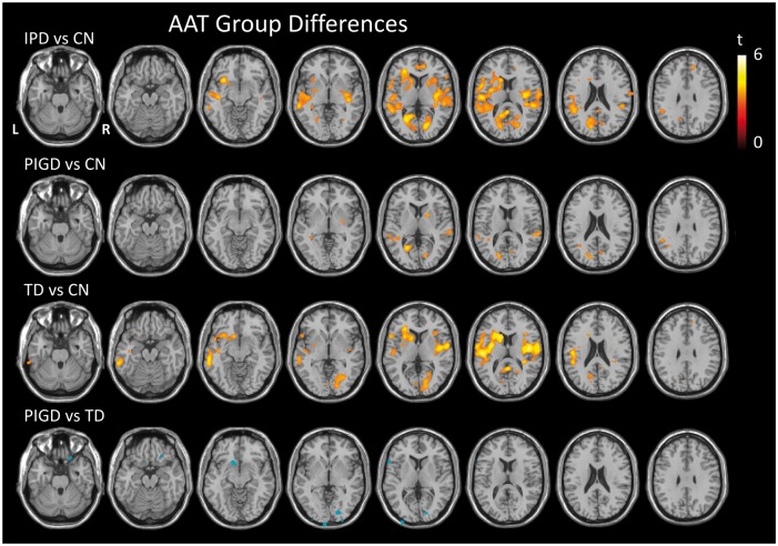
Figure 3.
Prolonged AAT in IPD compared to controls. T-statistic maps showing regions of significantly longer (red) and shorter (blue) AAT between pairwise group comparisons as indicated, thresholded at p < 0.001 uncorrected, minimum cluster size 50 voxels. AAT: arterial arrival time; IPD: idiopathic Parkinson’s disease.

