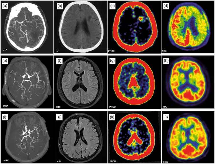
Figure 1.
Upper row: CTA in a 64-year-old man revealed severe stenosis of the left middle cerebral artery (MCA) (a), brain CT showed left frontal lobe infarction (b). PET/CT scan showed accumulation of 68Ga-NOTA-PRGD2 around the infarct area (c) with a diffuse decrease in the metabolism (yellow arrow) in 18F-fluorodeoxyglucose (FDG) PET (d) 33 days after ischemic stroke. Middle row: A 44-year-old man had an ischemic stroke 72 months prior to enrolment, leaving numbness in his left arm. MRA revealed right MCA occlusion (e). No significant infarcts were seen on the MRI (f). The 68Ga-NOTA-PRGD2 uptake was found in a puncta-like form scattered over the subcortical areas (g, yellow arrow), although no significant difference for 18F-FDG PET was seen between the lesioned and contralateral sides (h). Lower row: A 70-year-old man had an ischemic stroke 22 years prior to enrolment, leaving weakness in the right leg. MRA showed left MCA occlusion (i). No infarct was observed on the MRI (J). The 68Ga-NOTA-PRGD2 PET and 18F-FDG PET uptakes were not significantly different between the lesioned and contralateral sides (k, l).
CTA: computed tomography angiography; PET: positron emission tomography; CT: computed tomography; MRI: magnetic resonance imaging; MRA: magnetic resonance angiography.

