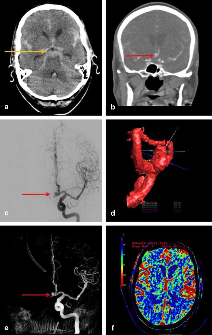
eFigure 1:
Imaging studies for the detection of subarachnoid hemorrhage and the determination of the source of bleeding.
These are the initial imaging studies obtained for a 63-year-old man who was admitted because of the sudden onset of the worst headache of his life, followed by a decline in consciousness. A computed tomogram (CT) of the head displays the classic pattern of a basal subarachnoid hemorrhage, with blood in the basal cisterns appearing as a hyperintense signal in the pentagonal cistern (image a, orange arrow). Subsequent CT angiography and digital subtraction angiography (images b, c) reveal an aneurysm of the anterior communicating artery as the source of bleeding (red arrows). The corresponding 3D reconstructions (images d and e) reveal the spatial configuration of the aneurysm and its spatial relationships to the nearby vessels. A perfusion CT (image f) enables visualization of cerebral perfusion: the parameter displayed here is the regional cerebral blood flow (rCBF)

