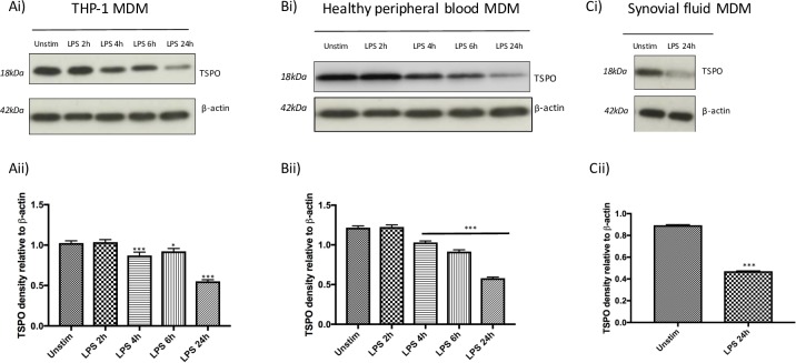
Fig 6. Western blotting indicates TSPO protein expression decreases on treatment of MDM with M1 stimuli LPS plus IFN-γ.
(i) representative western blot of TSPO protein with β-actin acting as loading control and (ii) densitometry normalized to β-actin in (A) THP-1 MDM, (B) healthy peripheral blood MDM and (C) synovial fluid MDM either unstimulated (unstim) or treated with 10ng/mL LPS plus 20ng/mL IFN-γ (LPS) for 2,4,6 and 24 hours. Densitometry data is expressed as the mean of five independent experiments ± SEM. (*p<0.05, **p <0.01, ***p≤0.001).

