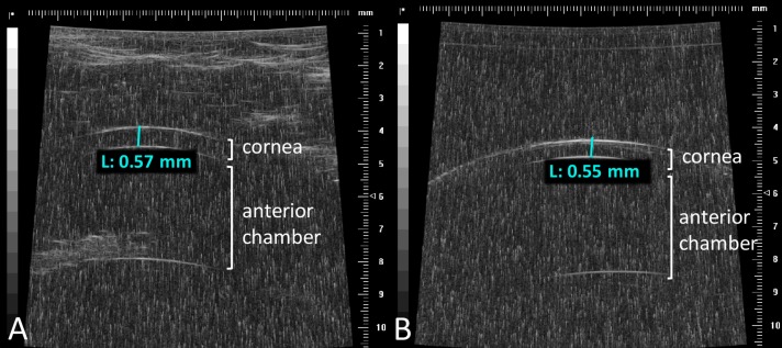
Figure 5.
Ultrasound measurement of lamb cornea before and after a 1-hour exposure to 1000 ppm NO. Representative ultrasound images of the ocular anterior segment, obtained in lambs (A) before and (B) after topical eye exposure to 1000 ppm NO for 1 hour. The corneal thickness was similar (A) before and (B) after exposure to 1000 ppm NO. The cornea, anterior chamber, and the iris are evident. Depth of the cornea did not differ in NO-treated versus control-gas treated eyes.

