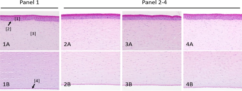
Figure 6.
H&E staining of lamb corneas before and after exposure to 1000 ppm NO. Panel 1: control animal. Panels 2–4: NO-treated animals. (A) Corneal epithelium and anterior stroma. (B) Posterior stroma, Descemet's membrane, and endothelium. No microscopic lesions or differences between groups were detected. H&E, ×20 objective. [1] Epithelium; [2] Bowman's membrane; [3] Stroma; [4] Descemet's membrane.

