. 2017 Sep 11;6:e28275. doi: 10.7554/eLife.28275

Figure 1. Uniform Bcd expression specifies cell fates corresponding to levels of expression.

(A) Wild-type, bcd null mutant (bcdE1), and maternal hunchback, nanos, torso-like (hb nos tsl) triple mutant and bcd hb nos tsl mutant embryos at NC14 immunostained with antibodies against Bcd, Btd, and Kni. Embryos are oriented with anterior to the left. The anterior Kni domain (filled arrow) is absent in bcd but restored in hb nos tsl embryos, while the posterior stripe (open arrow) shifts anteriorly in in bcd but expands posteriorly in hb nos tsl. Neither Btd nor Kni exhibit patterned expression in bcd hb nos tsl. Images are maximum z-projections and image contrast was adjusted uniformly across the entire image for display. See Figure 1—figure supplement 1A for quantification of Kni intensity between genotypes. (B) Expression levels of uniform GFP-Bcd transgenic constructs relative to wild-type Bcd expression. Live embryos were imaged in during NC14, and dorsal profiles were plotted. Error bars are standard error of the mean. For wild-type, n = 23 embryos; bcd-uBcd n = 13; mtrm-uBcd n = 7; and αTub67C-uBcd n = 14. See also Figure 1—figure supplement 1D and Table 6. (C) Immunostaining as (A), for each level of uniform Bcd. Anterior target gene expression is absent at the lowest level. At intermediate (mtrm) and high (αTub67C) levels of uBcd, anterior expression patterns are expanded and/or duplicated in the posterior, and posterior expression of Kni is absent. (D) Larval cuticle preparations for the indicated genotypes. Embryos are oriented with anterior at the top. Head structures are indicated with open arrows and tail structures with filled arrows. αTub67C >uBcd embryos develop essentially no cuticle tissue, but form only what appear to be anteriorly-derived mouth structures. mtrm >uBcd results in a duplication of the anterior-most abdominal denticles in the anterior and posterior of the embryo, with no clear terminal structures forming at either end. bcd >uBcd embryos have a normal posterior and all abdominal segments, but no thoracic or head structures. Images of individual embryos were rotated and cropped to exclude nearby embryos and air bubbles.

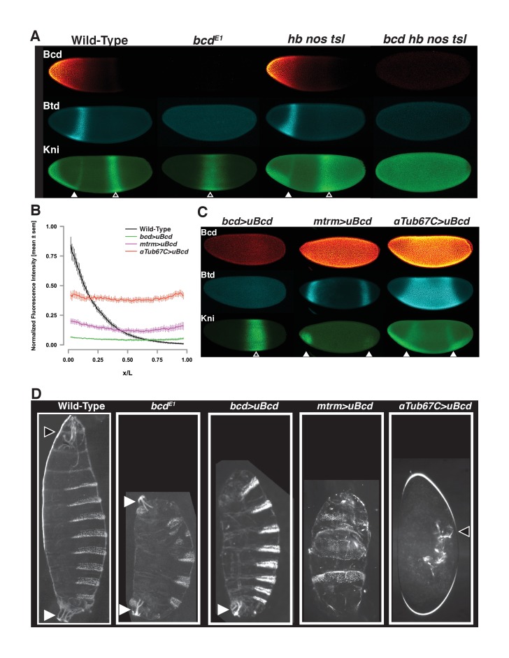
Figure 1.

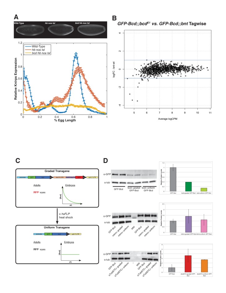
Figure 1—figure supplement 1. Target gene expression and Bcd binding in maternal matterning mutants and features the uniform Bcd transgene.

Figure 1—figure supplement 1.

(A) Quanitification of Knirps intensity in wild-type, triple mutant (hb nos tsl) and quadruple mutant (bcd hb nos tsl). Bicoid activates patterned expression of Knirps. In embryos in which Bicoid is the only source of maternal patterning information (hb nos tsl), a broad domain of Kni is expressed in the posterior of the embryo. In quadruple mutant embryos, a low level of uniform Knirps is expressed ubiquitously, suggesting that Bcd is required for activating expression of knirps above a background level. Heat-fixed embryos from wild-type (Oregon-R) mothers, hunchback nanos torso-like germline clones and bicoid hunchback nanos torso-like germline clones were pooled and immunostained in a single tube with a rat anti-Knirps primary antibody and Alexa-647 rat antibody. Embryos were mounted on a single slide and imaged by confocal microscopy. Representative embryos for each genotype are shown. Fluorescence intensity of Knirps was extracted from dorsal profiles of midsagittal sections of embryos and plotted using MATLAB. Data are fluorescence intensity minus background, and error bars are standard error of the mean for n = 5 wild-type, n = 8 hb nos tsl, and n = 6 bcd hb nos tsl embryos. (B) Smear plot generated in EdgeR (Robinson et al., 2010) showing the log transformed fold-change in Bcd binding between mutant and wild-type embryos for each Bcd peak, vs. the average log transformed sequencing read counts per million (CPM). Bcd binding shows no significant changes between wild-type and nos tsl mutant embryos. Significance was determined using EdgeR to perform a pairwise exact test with a cutoff of FDR ≤ 0.05, comparing binding between eGFP-Bcd;;bcdE1 and eGFP-Bcd;; bcdE1 hbFB nosL7 tsl4 in the 1,027 Bcd peaks. (C) Schematic of the uniform Bcd transgene. The uniform Bcd transgene contains an N-terminal GFP-tagged Bcd driven by the various maternal promoters discussed in the text. Downstream of the bcd coding sequence is a cassette containing the endogenous bcd 3'UTR and a 3xP3-hsp70 promoter driving promoter of RFP. This cassette is flanked by FRT sites. The sqh 3'UTR lies downstream of the FRT cassette. Flies expressing this version of the transgene can be identified by RFP expression in their eyes, and females produce embryos in which Bcd is distributed in a gradient. Males from this transgenic stock are crossed to females expressing a heat shock inducible flippase (hsFLP), and heat shocking the F1 larvae results in recombination and excision of the cassette at the FRT sites, bringing the sqh 3'UTR directly downstream of the bcd coding sequence. This initially results in mosaic F1 flies with a mosaic graded/uniform Bcd germline. The F1 are further outcrossed to bcdE1 mutants and F2 individuals producing embryos with uniform Bcd distributions can be identified by the lack of RFP expression in the eyes. (D) Expression levels of uniform Bcd constructs measured by western blots. Western blots for GFP-Bcd were performed on embryos at NC14. Representative gels and quantifications are shown for the bcd promoter-driven transgene (A), mtrm promoter-driven transgene (B) and αTub67C promoter-driven transgene (C). In the barplots, band intensities are reported relative to wild-type (GFP-Bcd). All lanes are normalized to an α-tubulin loading control. Error bars are standard deviation between two biological replicates for each sample. MW = molecular wt marker, *=skipped lane.