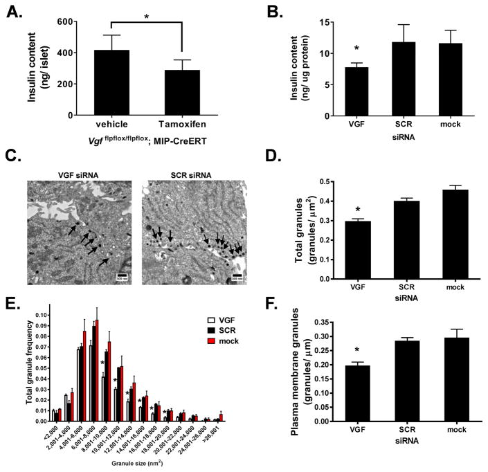
Figure 3. VGF suppression reduces secretory granule number and size.
(A) Insulin content was determined from islets isolated from control (vehicle) and β-cell VGF KO (tamoxifen; Tm) mice. (B–F) 832/3 cells were transfected with rat VGF siRNA duplexes (SMARTpool), non-targeting control duplex (SCR), or mock transfected as indicated. (B) Insulin content was determined from whole cell lysates. (C–F) 832/3 cells were treated with 2.5 mM Glc for 1 hr followed by 12 mM Glc for 30 minutes and processed for electron microscopy. (C) Representative electron micrographs with arrows indicating dense core granules. (D) Total number of dense core granules normalized to total cell area. (E) Frequency distribution of binned total granule sizes. (F) Number of granules within 200 nm of the plasma membrane normalized to linear plasma membrane surface. Data represent the mean ± S.E.M of n = 6 mice per group (A) or 3 independent experiments (B, D–F). * p ≤ 0.05 as compared to control (vehicle) islets (A, unpaired ttest); or SCR siRNA- or mock-transfected cells (B, D, F; one-way ANOVA, Tukey post-test; E, two-way ANOVA, Bonferroni post-test ).

