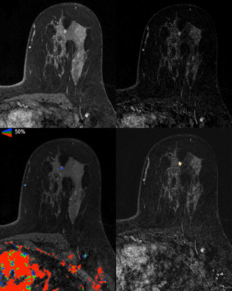
Figure 1.

Axial images of A) T1 fat-suppressed fourth post-contrast sequence, B) subtracted fourth post-contrast sequence, C) iCAD color registration of the subtracted first post-contrast subtraction sequence, and D) 3D Slicer segmentation of the subtracted fourth post-contrast sequence showed a 6 mm irregular mass in the left upper breast of a 36-year-old woman with a known contralateral invasive cancer (not shown); excision showed ER positive and HER2 negative intermediate-grade DCIS.
