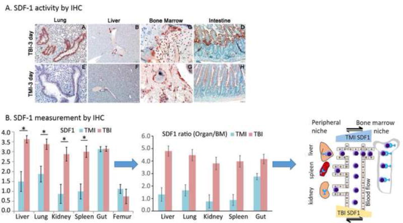
Figure 5.

Intestinal tissue damage assessment and marrow and tissue SDF-1 expression in mice receiving TBI versus TMI, 3 days after HCT. A. Intestinal histology and measured villus height and crypt depth. B. Tissue SDF-1 measurement by IHC is presented for various organs. The derived ratios of SDF-1 in organs with respect to bone marrow are also presented. A schematic presentation showing, for TBI, increased SDF-1 chemokine gradient towards the peripheral niche compared to the BM niche, attracting migratory donor cells from blood to organs; for TMI the reverse migration occurs, with higher chemokine gradient towards the BM niche.
