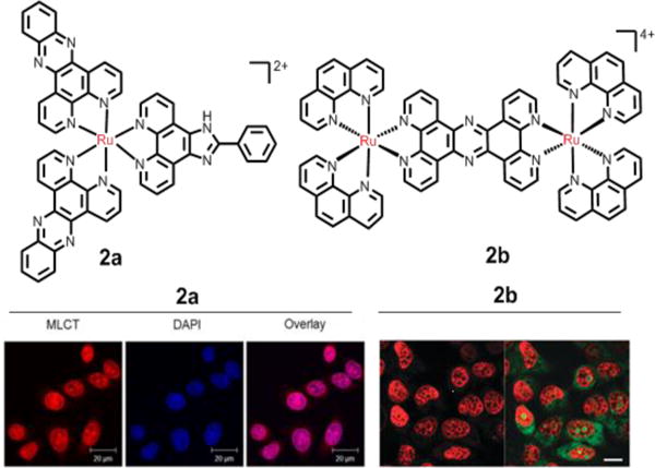
Fig. 5.

Representation of Ru(II) compounds that target DNA. Left-hand fluorescence image: the nuclear DNA staining of 2a in PFA-fxed HeLa cells, as evident by red luminescence, with co-staining by nuclear DNA dye DAPI. Right-hand fluorescence image: the nuclear DNA staining of the dinuclear 2b in MCF-7 cells, as evident by the red luminescence, with co-staining SYTO-9 (green). Reproduced with permission from ref. 109 and 111, respectively. Copyright 2015, Nature Publishing Group.
