Figure 7.
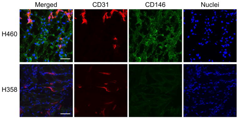
CD146 and CD31 co-staining of lung tumor sections from H460 and H358 tumor-bearing mice. CD146 staining (green) was elevated in H460 sections, while only background signal was visible in H358 sections. CD31 staining (red) revealed similar degrees of vasculature between both tumor models. Scale bar = 100 μm.

