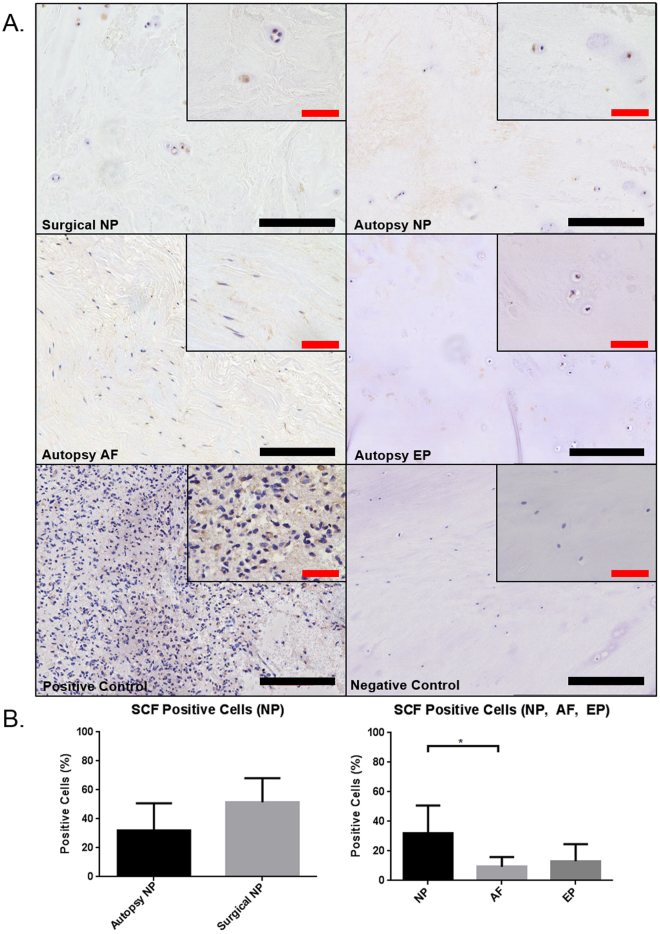
Figure 2.
Immunohistochemical staining for mast cell chemoattractant stem cell factor (SCF) in human IVD (SCF) (A). The positive control was tissue taken from human brain tissue. Quantification of percent positive cells for SCF demonstrated no significant differences between surgical and autopsy specimens (p = 0.081) however regional differences were observed between NP and AF (B) (p = 0.017) (Autopsy N = 7, Surgical N = 6). Black scale bar = 200 µm, Red scale bar = 50 µm.

