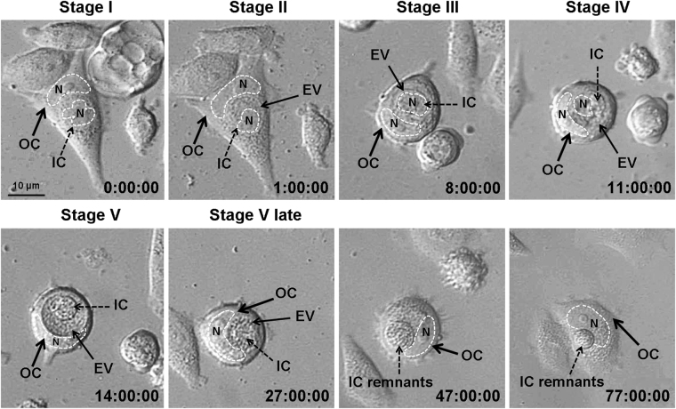
Figure 3.
Representative time-lapse imaging of entosis in a MCF7 cell monolayer. Shown is the full sequence of events from cell penetration into another cell and up to its degradation. IC, inner cell; OC, outer cell; EV, entotic vacuole. Nuclei (N) are encircled by the dashed lines. In 9 out of 13 examined cells, the inner cells were degraded at the end of entosis, while in the remaining 4 cells the inner cell exited the entotic vacuole.

