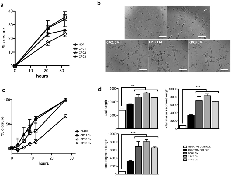
Figure 4.
Functional evaluation of the CPC secretome. (a) CPC wound healing capacity. All CPC isolates (CPC1-3) showed enhanced repair capacity compared with HDF cells; expressed as the percentage of scratch closure. (b) HDF cells in CM from the three CPC isolates showed improved repair capacity compared with the negative control (DMEM); expressed as percentage of closure. (c,d) Angiogenic activity in CM from the three CPC isolates compared with negative (C+) and positive controls (C+), using HUVEC (see Methods); after 6 h incubation, tube formation was analyzed using ImageJ software. Bar, 500 μm. Data are expressed as mean ± SD; n = 3.

