Figure 1.
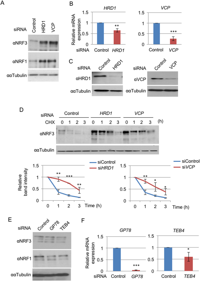
Hrd1 and VCP regulate the cytoplasmic degradation of NRF3. (A) The addition of HRD1 or VCP siRNA stabilized endogenous NRF3 in DLD-1 cells. At 48 hr after the siRNA transfection, the whole-cell extracts were prepared and analyzed by immunoblotting with anti-NRF3 and anti-NRF1 antibodies. α-Tubulin was used as an internal control. (B,C) The knockdown efficiency of HRD1 and VCP siRNA was determined by quantitative real-time PCR (qRT-PCR) analysis and immunoblot analysis with anti- HRD1 and anti-VCP antibodies. The values of the qRT-PCR analysis in (B) were normalized to the 18S rRNA data. (D) The knockdown of HRD1 or VCP inhibited NRF3 degradation in the cycloheximide chase experiment. The immunoblot analysis was performed with the anti-NRF3 antibody. α-Tubulin was used as an internal control. The graphs depict the quantified band intensities of NRF3, normalized to that of α-Tubulin. (E) The addition of GP78 or TEB4 siRNA did not stabilize the endogenous NRF3 in DLD-1 cells. The experiment was done as described in the legend of Fig. 1A. Error bars (B,D and F) represent data from three independent experiments (mean ± standard deviation). The two-tailed Student’s t-test was used for the statistical analysis. *P < 0.05, **P < 0.01 and ***P < 0.001 compared to the Control data.

