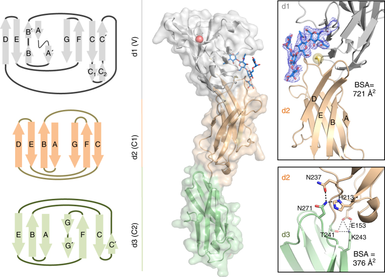
Fig. 1.
Three-dimensional structure of human CD22. Crystal structure of the three N-terminal Ig domains of human CD22 (d1–d3) represented as a schematic diagram, secondary structure cartoon, and transparent surface. d1 (gray) adopts a V-type Ig domain fold and R120 known to bind Sia is depicted as a red sphere. d2 (wheat) adopts an unexpected C1-type Ig domain fold, while d3 (green) is of C2-type fold. Insets show inter-domain interfaces of d1/d2 (top) and d2/d3 (bottom). CD22 d2 strands D–E are unusually elongated and contribute to the extensive d1–d2 interface. A blue mesh represents the composite omit electron density map (1.0 σ contour level) associated with the N101 glycan (sticks) involved in shaping the d1/d2 interface. Inter-domain disulphide bond C39-C167, highly conserved among Siglec family members, is shown as a yellow sphere

