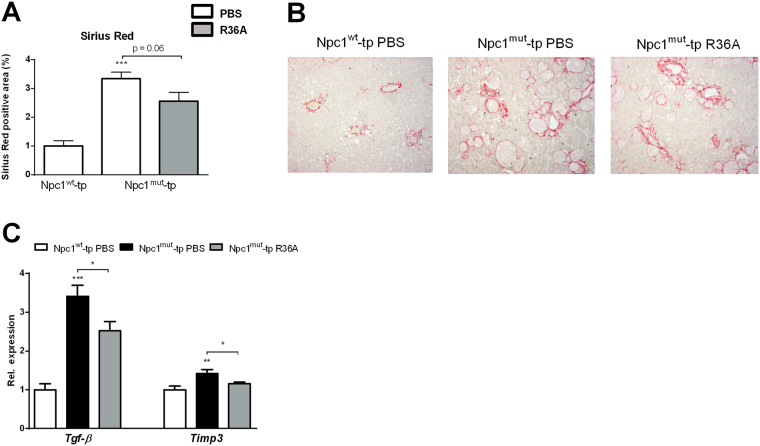
Figure 5.
Parameters of hepatic fibrosis. (A) Quantification of Sirius Red (collagen) staining. (B) Representative pictures of Sirius Red staining (original magnification, 100x) of Npc1 wt-tp mice and Npc1 mut-tp mice with or without immunization on an HFC diet for 3 months. (C) Gene expression analysis of the fibrosis markers, Tgf-β and Timp3. n = 9–11 mice/group. Gene expression data are shown relative to Npc1 wt-tp mice by use of two-tailed unpaired t test. *p < 0.05; **p < 0.01; ***p < 0.001. All error bars are SEM.

