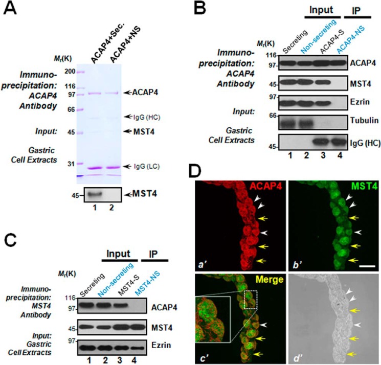
Figure 1.
ACAP4 is a novel MST4 interaction partner in histamine-stimulated gastric parietal cells. A, ACAP4 forms a novel complex with MST4 in secreting parietal cells. Aliquots of secreting (histamine-treated) and NS gastric parietal cells were collected by centrifugation and extracted with Triton X-100–containing buffer as described under “Experimental procedures.” Clarified cell lysates were incubated with Sepharose bead affinity matrix covalently coupled with ACAP4 mouse antibody as described under “Experimental procedures.” The beads were washed successively with PBS before elution with 0.2 m glycine (pH 2.3). All samples were separated by SDS-PAGE. The ACAP4 protein band migrating at 99 kDa along with the 45-kDa bands were then removed for mass spectrometric identification. Western blot analysis confirmed that the 45-kDa band is MST4 (bottom panel). HC, heavy chain; LC, light chain. B, ACAP4 immunoprecipitates (IP) from secreting and non-secreting gastric parietal cells were immunoblotted with antibodies against ACAP4, MST4, ezrin, and tubulin. Note that only ACAP4 immunoprecipitates from secreting parietal cells brought down ezrin and MST4 but not tubulin. Neither ezrin nor MST4 were observed in ACAP4 immunoprecipitates from non-secreting parietal cells. C, MST4 immunoprecipitates from secreting and non-secreting gastric parietal cells were immunoblotted with antibodies against ACAP4, MST4, and ezrin. Note that only MST4 immunoprecipitates from secreting parietal cells brought down ACAP4. D, isolated gastric glands were labeled with antibodies against ACAP4 (a′, red), MST4 (b′, green) and imaged for phase contrast (d′). Images from ACAP4 (a′, red) and MST4 (b′, green) were merged to assess their co-distribution profiles in gastric glandular cells (c′). Scale bar = 10 μm. Note that ACAP4 is co-localized to the corresponding MST4-positive gastric parietal cells (white arrowheads) but not non-parietal cells (yellow arrows).

