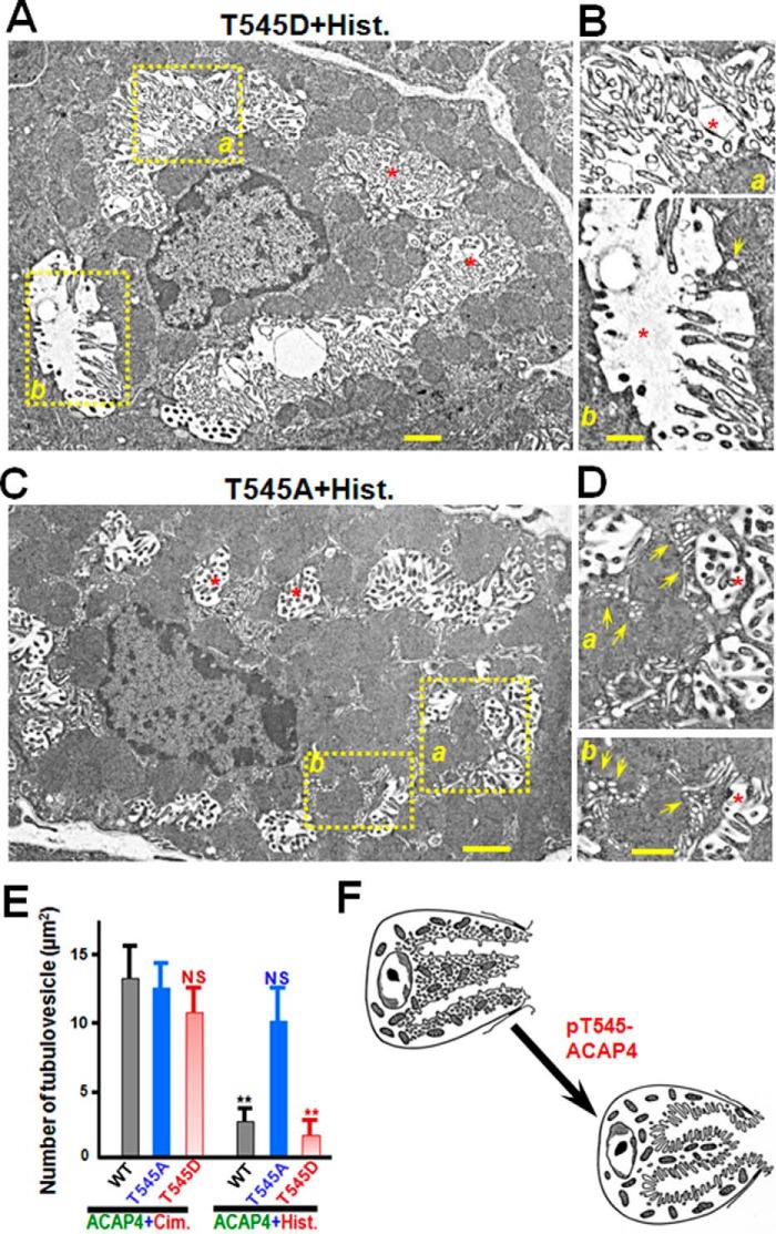
Figure 5.
Phosphorylation of ACAP4 by MST4 enables recruitment of tubulovesicles to the apical membrane of parietal cells. A, in the resting cell, short apical microvilli are supported by long microfilaments extending deep into the cytoplasm. Stimulation leads to docking and fusion of H,K-ATPase–rich tubulovesicles to the apical plasma membrane, greatly expanding the apical surface. Interactions between the apical membrane and microfilaments, via ezrin–ACAP4 interaction, were postulated to reorganize the expanded surface into long microvilli. Aliquots of cultured parietal cells were transiently transfected to express GFP–ACAP4T545A and GFP–ACAP4T545D mutants, followed by histamine stimulation. The preparations were processed for classic transmission electron microscopic analyses as described under “Experimental procedures.” Thin sections were post-stained with uranyl acetate and lead citrate and examined under an electron microscope. In histamine-elicited secreting parietal cells expressing ACAP4T545D, the greatly expanded apical membrane is lined with numerous elongated microvilli. Scale bar = 1 μm. B, magnified view of A (insets, a and b) showing the elongation of microvilli in proximity to the microvillar membrane in parietal cells with concurrent diminished tubulovesicles. Only a few tubulovesicles can be found after careful examination (b, arrow). Scale bar = 500 nm. C, in histamine-treated parietal cells expressing ACAP4T545A, short apical microvilli were evident as long microfilaments extending deep into the cytoplasm, reminiscent of non-secreting cells. Scale bar = 1 μm. D, magnified view of C (insets, a and b) showing the abundance of tubulovesicles in proximity to the microvillar membrane in parietal cells with concurrent minimized apical canalicular expansion (*). Dozens of tubulovesicles were readily apparent in parietal cells expressing ACAP4T545A (a and b, arrows). Scale bar = 500 nm. E, quantitative analyses of tubulovesicle density in parietal cells expressing ACAP4T545A and ACAP4T545D, respectively. Error bars represent S.E.; n = 10. **, p < 0.01; NS, no significant difference. Cim., cimetidine; His., histamine. F, schematic illustrating the role of phosphorylation of ACAP4 at Thr545 in promoting the fusion of tubulovesicles to the apical membrane of parietal cells upon MST4 activation by histamine.

